What Is the Significance of Mosaic Embryos?
With the advent of chromosomal testing of blastocysts, we began to recognize that a significant percentage of embryos have abnormal chromosomes. This is an obvious explanation for a pregnancy rate <100% when transferring embryos and an IVF cycle.
In addition, this has been an explanation for the decrease in fertility as women age as the percentage of embryos with abnormal chromosomes goes up with age. This is not unexpected since we know that in women who have a miscarriage, chromosomal testing on the fetal tissue is more likely to be abnormal as women age. In addition, the frequency of babies born with chromosomal abnormalities such as Down’s syndrome goes up significantly with age of the mother.
When PGS-A testing was first performed, embryos were classified as euploid or normal meaning that the cells had normal chromosomal numbers or aneuploid or abnormal meaning that there were abnormal cells in the embryo. The "experts" said that embryos with normal chromosome testing should be made available for transfer; however, embryos with abnormal chromosomes should NEVER be transferred. The concern was that these abnormal aneuploid embryos could result in the birth of an abnormal baby.
Over time, the designation of embryos has changed. We now talk about normal euploid embryos, low-level mosaic embryos (LLM), high level mosaic embryos (HLM) and abnormal aneuploid embryos. Again the "experts" suggested that the mosaic embryos not be transferred because of the fear of creating a baby with mosaicism. Nevertheless, there have been several programs in the country who decided that transfer of mosaic embryos was acceptable and to many experts’ surprise, normal pregnancies and babies sometimes resulted.
When I first saw these reports, I had to stop and remind myself that prior to our ability to test the chromosomes on embryos before embryo transfer, we transferred all the embryos that a patient would make. The pregnancy rate especially in older women was low but none of the babies born had mosaicism. This is even though I am sure that many abnormal and mosaic embryos were being transferred without our knowledge because the chromosome testing on the embryos had not been performed. It was at that point that we decided at Denver Fertility Care that we would consider and even encourage the transfer of mosaic embryos especially in patients who had no normal embryos and had only mosaic embryos.
Shortly after that, I was at a national fertility meeting and during a panel discussion regarding mosaic embryos there was a discussion on the pros and cons of transferring mosaic embryos. After the expert against transferring mosaic embryos spoke, I got up and asked the question as to whether he had ever transferred a mosaic embryo. He told me absolutely never. I very graciously stated that I did not believe him. You can only imagine the flurry of conversation in the conference room. I then asked him if he had been performing IVF treatments prior to the advent of chromosome testing of embryos. He said yes. I stated that in that case I am sure that he had unknowingly transferred many abnormal as well as mosaic embryos in the past. I asked him if he had ever had a baby born with mosaicism. He said no, but he had several patients who ended up with babies with Down’s syndrome. And this is not to be unexpected with untested embryos.
What is a Mosaic Embryo?
The best analogy of mosaicism is looking at a mosaic tile floor made up of 2 colors of tiles, red and green. (See the Figure) Green tiles are normal and give us a go ahead to perform a transfer and expect a normal pregnancy. Red tiles are abnormal and suggest that if a transfer is performed, an abnormal pregnancy will take place. With a low-level mosaic pattern (LLM), 20-50% of the tiles may be red tiles and the remaining 50-80% are green tiles. With a high-level mosaic pattern (HLM), 50-80% of the tiles are red and the remaining 20-50% are green.
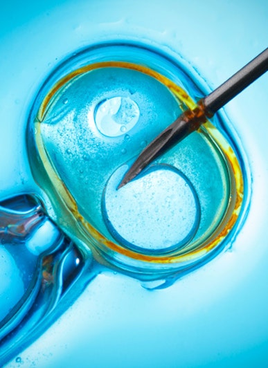
When we perform PGT-A (preimplantation genetic testing for aneuploidy), the portion of the embryo that is biopsied to test is the trophectoderm that will form the placental cells. The inner cell mass are the cells that form the actual baby. We should biopsy the inner cell mass to have the true reflection of the future baby, but everyone is afraid of disrupting the inner cell mass and thereby possibly disrupting the chances for the embryo to become a pregnancy and baby. Moreover, there is a high concordance between the chromosomes in the trophectoderm and the inner cell mass.
When the PGS (preimplantation genetic screening) biopsy comes back showing mosaicism, the question that remains unanswered is whether the inner cell mass and therefore the baby also shares this mosaicism or if the inner cell mass is normal.
We know that embryos are remarkable in that if the embryo detects abnormal cells, it can segregate those cells away from the inner cell mass either by "kicking" them into the trophectoderm or completely out of the embryo. This self-policing by embryos allows the embryo to continue normal development rather than continuing as an abnormal embryo. The trophectoderm and eventually the placenta itself can tolerate abnormal cells and still function as a viable pregnancy.
An embryo with a mosaic result is at increased risk of having the inner cell mass also be mosaic. An embryo with the LLM result has fewer abnormal cells than an embryo with HLM. The likelihood for the inner cell mass to be abnormal is therefore going to be higher with a HLM embryo than with an LLM embryo and consequently the chances for no pregnancy or a miscarriage will be higher with an HLM embryo.
The data is still accumulating; however, studies would suggest that LLM embryos may have an ongoing pregnancy rate as high as 40-50% but as many as half of these pregnancies may go on to miscarry because of aneuploidy in the fetus. Of the pregnancies that do not miscarry, most will have normal testing by amniocentesis. The data set is not large enough to state that every ongoing pregnancy will be normal and hence the strong recommendation for an amniocentesis for genetic testing. With HLM embryos, the ongoing pregnancy rate may be as high as 20-25% but again as many as half of these pregnancies will miscarry because of abnormal chromosomes in the fetus.

Which Mosaic Embryos Should Be Used for Transfer?
When the laboratory gives us information on abnormal or mosaic embryos, they can tell us which chromosomes are abnormally represented and whether there is an extra chromosome or a missing chromosome. There are 22 pairs of autosomal (nonsex) chromosomes and 1 pair of sex chromosomes either XX or XY depending upon whether the embryo is female or male. Most chromosomal abnormalities are lethal to the embryo, and the embryo probably dies shortly after it is transferred to a woman's uterus. Other chromosomal abnormalities are less lethal, and a pregnancy might ensue, but because the chromosomal abnormality is ultimately lethal, a miscarriage occurs.
Rare chromosomal abnormalities such as sex chromosome abnormalities and chromosomes 21, 18, 16 and 13 can be born alive.
The sex chromosomal abnormalities are less severe than those caused by autosomal chromosomes. Sex chromosome abnormalities include Turner’s syndrome (45, XO), super female (47, XXX), Klinefelter's syndrome (47, XXY) and 47, XYY. Patients with Turner’s syndrome and Klinefelter's syndrome are generally infertile. The other sex chromosome abnormalities may have some reduction in fertility but can naturally have children.
The autosomal chromosome abnormalities are generally more severe. Trisomy 18 or Edwards’ syndrome is usually fatal before birth and the few that are born alive usually die within the first year of life. There have been reports of trisomy 18 mosaicism in babies and they generally have a better prognosis.
Trisomy 16 is the most common autosomal trisomy in humans but is almost uniformly embryonic lethal. Usually, death occurs by the 10th week of pregnancy. It is rare for these babies to survive into the third trimester and if born alive die shortly after birth. It could be possible for a mosaic trisomy 16 fetus to survive; however, there are no reports of mosaic trisomy 16 babies.
Trisomy 13 or Patau syndrome is usually lethal during mid pregnancy, but some babies can be born alive. They usually die before the end of their first year.
The other autosomal trisomies are uniformly lethal either at the embryo stage resulting in no pregnancy or within the first trimester of pregnancy resulting in a miscarriage. Babies with mosaicism of these other autosomal trisomies have not been reported but certainly could theoretically occur.
Autosomal monosomies are extremely rare. Monosomies of the larger chromosomes (1-13) or gene-dense chromosomes (17 and 19) do not survive; however, in the mosaic state there have been reports of births. There also are a few reported cases of autosomal monosomies of the smaller chromosomes (14, 15, 16, 18, 20, 21 and 22) surviving but these babies have severe physical and developmental sequelae and usually die shortly after birth. Again, in the mosaic state, there may be a higher likelihood of milder symptoms and survival.
I have often thought that it would be extremely informative if every human being had chromosomal testing to determine their personal chromosomal status. It could be that many people who have minimal physical or developmental abnormalities have unrecognized mosaicisms. It is important to realize that this is purely my conjecture as there is no data to suggest that these people actually do exist.
There are numerous reports in the literature of sex chromosome mosaicism and of trisomy 21 mosaicism.
While on the one hand, the likelihood of a mosaic embryo resulting in the birth of an abnormal child with mosaicism is low, I recommend that all couples who have a pregnancy after the transfer of the mosaic embryo proceed with genetic counseling and diagnosis in pregnancy. Because the mosaicism of the embryo was diagnosed from a biopsy of the trophectoderm which is the part of the embryo that becomes the placenta, performing the NIPT testing in early pregnancy or a chorionic villous sampling may not be the optimal test to determine that the pregnancy is normal or abnormal as both of these tests are actually testing cells from the placenta. If either of those tests are performed and are normal, the likelihood for an abnormal baby is reduced; however, an amniocentesis is the form of testing that we would recommend to confirm that the pregnancy is truly normal.


